
Fig. 1
- ID
- ZDB-FIG-170605-5
- Publication
- White et al., 2017 - Immunomodulation-accelerated neuronal regeneration following selective rod photoreceptor cell ablation in the zebrafish retina
- Other Figures
- All Figure Page
- Back to All Figure Page
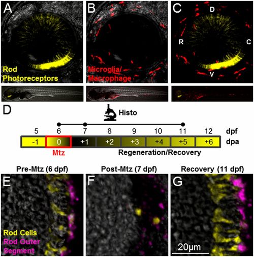
Intravital imaging and prodrug-induced cell ablation. (A–C) Whole-retina z-projection images of double-transgenic (rho:YFP-NTR;mpeg1:Tomato) 6-dpf larvae in which rod cells express YFP-NTR (A and C, yellow), and macrophage/microglia express tdTomato (B and C, red). The upper images show the whole eye (34× magnification); the lower images show the whole fish (4× magnification). (D) Schematic of the assay and analysis timeline for data shown in E–G. Retinal sections were processed for immunohistology (Histo) and imaged at time points indicated by dots on the Histo line. Mtz exposures occurred over a 24-h span at 6 dpf (red box) with loss and subsequent rod cell regeneration (black/yellow gradient) occurring over a weeklong recovery period (−1 to +6 dpa). (E–G) Retinal sections from a rho:YFP-NTR transgenic larva treated with Mtz (10 mM) for 24 h, starting at 6 dpf (E), demonstrating ablation (F), and subsequent regeneration (G) of rod photoreceptors indicated by the loss and recovery of YFP-expressing rod cells (yellow) and a rod outer segment marker (magenta, monoclonal antibody zpr-3, also known as “Fret 11”). C, caudal; D, dorsal; R, rostral; V, ventral. |