
Fig. S4
- ID
- ZDB-FIG-170601-21
- Publication
- Pietri et al., 2017 - The Emergence of the Spatial Structure of Tectal Spontaneous Activity Is Independent of Visual Inputs
- Other Figures
- All Figure Page
- Back to All Figure Page
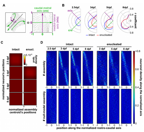
The neuronal assemblies are distributed along the caudo-rostral axis, related to Figure 3. (A) Example of normalization of the neuronal coordinates in a common spatial reference map, where each dot represents a neuron in its original position (left) and after the normalization (right); the morphological medio-lateral (mla) and caudo-rostral axis (cra) are represented in green and purple, respectively. (B) Correlation coefficients between the neurons belonging to each assembly and their respective centroids along different axes of the common spatial reference map, rotated every 15° for a total of 180°, for each tectum and for all developmental stages, in intact (blue) and enucleated (red) larvae. Note that the highest correlations were obtained along the tectal caudo-rostral axis in all considered conditions. (C) Null-model density plots of the caudo-rostal normalized positions of each neuron against the normalized position of each neuronal assembly centroid, along the caudo-rostral axis of the tectum, in intact and enucleated larvae, at each developmental stage; 0 is the most rostral position and 1, the most caudal one (indicated in the enucleated 8 dpf larva density plot). (D) Distribution of the density of the neurons per assembly along the normalized caudo-rostral axis (the axis was normalized between -1 to 1, from lateral left to lateral right) at all developmental stages, in intact (n = 622, 800, 1101 and 864 assemblies for 2.5, 3, 5 and 8 dpf stage respectively) and enucleated larvae (n = 511, 768 and 688 assemblies for 3, 5 and 8 dpf stage, respectively) and their respective null models (built from 50 repetitions of each neuronal assembly). Each neuronal assemblies are represented on the y-axis. |