
Fig. S3
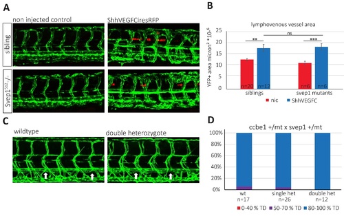
VEGFC over-expression and Svep1 and Ccbe1 interaction: (A – B) svep1 mutant endothelial cells respond to VEGFC. (A) Confocal projections of siblings and Svep1 mutants expressing VEGFC IRES RFP in the floorplate versus non injected control at 2 dpf, transgene: fli1a:GFP . Forced expression of human VEGFC in the floorplate led to excessive vessel sprouting both in siblings and in svep1 mutants (B) Quantification of endothelial vessel area as measured by GFP+ area surrounding a position of comparable RFP expression. Data sets were tested for normality (Shapiro-Wilk) and equal variance. P-values were determined by Student's t-test. Values are presented as means ± standard error of mean values (SEM). ns = not significant; * = P<0.05; ** = P<0.01; *** = P<0.001. (C - D) svep1 and ccbe1 do not genetically interact (C) Confocal projections of wt and svep1/ccbe1 double heterozygous animals do not show any defect in TD generation as compared to wildtype controls at 5 dpf, transgene: fli1a:GFP. Arrows indicate the position of the TD (D). Quantification of the extent of TD formation across ten body segments in the trunks of wildtype, single heterozygote and double heterozygote embryos do not indicate genetic interaction between svep1 and ccbe1. One out of 3 independent experiments is shown. |