
Fig. 3
- ID
- ZDB-FIG-170510-23
- Publication
- Chatzopoulou et al., 2017 - Functional analysis reveals no transcriptional role for the glucocorticoid receptor β-isoform in zebrafish
- Other Figures
- All Figure Page
- Back to All Figure Page
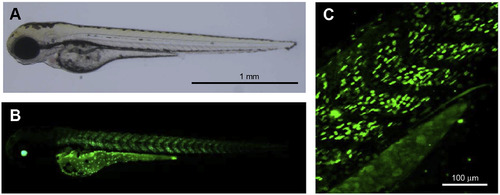
Images of a 3dpf zebrafish embryo generated by crossing the Tg(hsp70:Gal4) and Tg(UAS:GFP-zGRβ) transgenic lines. Embryos were heat shocked at 37 °C for 2.5 h at 1 and 2dpf. A. Transmitted light microscopy image showing representative embryo, indicating that transgenesis and heat shock treatment do not alter the morphology of zebrafish embryos. B. Fluorescence microscopy image showing expression of the GFP-zGRβ fusion protein. The green fluorescent signal is clearly present in cells in the muscles and the eyes, and in cells lining the yolk sac. C. Larger magnification fluorescence microscopy image showing expression of GFP-zGRβ in muscle tissue. The image shows that GFP-zGRβ is localized in the nuclear cell compartment. |
| Gene: | |
|---|---|
| Fish: | |
| Condition: | |
| Anatomical Terms: | |
| Stage: | Protruding-mouth |
Reprinted from Molecular and Cellular Endocrinology, 447, Chatzopoulou, A., Schoonheim, P.J., Torraca, V., Meijer, A.H., Spaink, H.P., Schaaf, M.J., Functional analysis reveals no transcriptional role for the glucocorticoid receptor β-isoform in zebrafish, 61-70, Copyright (2017) with permission from Elsevier. Full text @ Mol. Cell. Endocrinol.