
Fig. 7
- ID
- ZDB-FIG-170426-13
- Publication
- Li et al., 2009 - Medaka vasa is required for migration but not survival of primordial germ cells
- Other Figures
- All Figure Page
- Back to All Figure Page
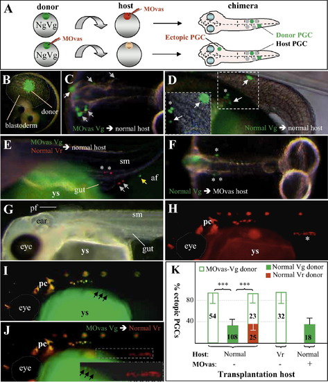
vasa controls PGC migration cell-autonomously by chimera assay. (A) Scheme of cell transplantation. Following zygotic MOvas injection, homozygous Vg donor blastula cells were transplanted into control non-transgenic (shown here) or transgenic Vr fish host blastulae. Donor PGCs (green) are daily monitored for normal (asterisks) and ectopic (arrows) distribution. In a reciprocal transplantation, non-transgenic embryos receive MOvas injection and used as the host to receive control Vg donor blastomeres. Ectopic PGCs are outside the migration route and gonad. (B–E) Transplantation into normal host. (B) Blastula immediately after transplantation showing approximately 50 GFP-positive donor cells among the host deep layer cells. (C) Chimera (2 dpf) from MOvas-injected donor, showing essentially all donor PGCs in ectopic sites. (D) Chimera (5 dpf) from transplantation between normal donor and host, showing normal as well as ectopic distribution of donor PGCs. (E) Chimeras (5 dpf) from cotransplantation of MOvas-injected Vg and non-injected Vr donors. RFP-positive PGCs from normal blastulae are seen both inside (asterisk) and outside (arrow) the gonad, whereas GFP-positive PGCs from vasa-depleted blastulae are outside in the anal fin (af). (F) Chimera (2 dpf) from reciprocal transplantation of normal Vg blastomeres into MOvas-injected non-transgenic hosts, showing normal distribution of vasa-depleted donor PGCs. (G–J) Chimera (7 dpf) by transplanting MOvas-injected Vg blastula cells into a normal Vr blastula. (G) Bright field micrograph. Clearly seen are many organs including the eye, ear, somites (sm), pectoral fin (pf) and gut. (H) Red fluorescent micrograph. Host PGCs are seen in the gonad. (I) Green fluorescent micrograph. Three vasa-depleted donor PGCs (arrows) are seen outside the gonad (insert). (J) Merged micrograph. The vasa-depleted donor PGCs do not colocalize with the host PGCs in the gonad. The insert shows the framed area at large magnification. Autofluorescent pigment cells (pc) and yolk sac (ys) are easily distinguishable from PGCs by morphology and location. (K) Statistical data of chimera assays. vasa knockdown of the donor (open column) is sufficient for ectopic PGC distribution regardless of whether the host environment is normal or vasa-depleted. For statistic analyses see the legend to Table 2. |
Reprinted from Mechanisms of Development, 126(5-6), Li, M., Hong, N., Xu, H., Yi, M., Li, C., Gui, J., Hong, Y., Medaka vasa is required for migration but not survival of primordial germ cells, 366–381, Copyright (2009) with permission from Elsevier. Full text @ Mech. Dev.