
FIGURE
Fig. 4
- ID
- ZDB-FIG-170425-6
- Publication
- Cui et al., 2017 - Chronic perfluorooctanesulfonic acid exposure disrupts lipid metabolism in zebrafish
- Other Figures
- All Figure Page
- Back to All Figure Page
Fig. 4
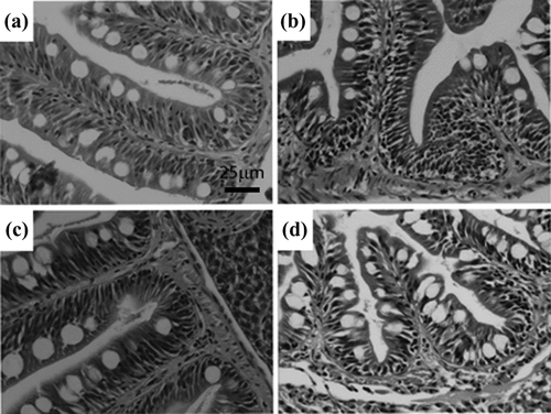
H&E staining of small intestine from F0 after exposed to PFOS for 180 days. Representative intestine sections from control female (a) and male (b), female exposed to 0.5 μM PFOS (c) and males exposed to 0.5 μM PFOS (d). Compared with the control males, the number of goblet cells was higher in PFOS-treated males, while the number of columnar cells decreased. There were no significant morphological changes in female following PFOS exposure. Scale bar: 25 µm; n = 5 fish per sex from each group. PFOS: perfluorooctanesulfonic acid; H&E: hematoxylin and eosin. |
Expression Data
Expression Detail
Antibody Labeling
Phenotype Data
Phenotype Detail
Acknowledgments
This image is the copyrighted work of the attributed author or publisher, and
ZFIN has permission only to display this image to its users.
Additional permissions should be obtained from the applicable author or publisher of the image.
Full text @ Hum. Exp. Toxicol.