
Fig. 2
- ID
- ZDB-FIG-170405-9
- Publication
- Stantic et al., 2015 - TAp73 suppresses tumor angiogenesis through repression of proangiogenic cytokines and HIF-1α activity
- Other Figures
- All Figure Page
- Back to All Figure Page
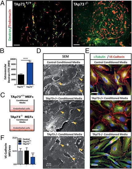
Loss of TAp73 increases tumor blood vessel permeability through reduced endothelial cell–cell contact. (A and B) TAp73+/+ and TAp73−/− tumors perfused with FITC-labeled dextran and stained for endothelial cells (endomucin). Green, FITC-labeled dextran leakage into the extravascular tumor space; red, endomucin staining of blood vessels. Mean fluorescent intensity (MFI) was determined for total dextran-FITC (green) signal with pixel counting; intravascular dextran-FITC (yellow) was subtracted from the final value (n = 5/group, five fields/tumor was used for quantification; ****P < 0.0001). (Scale bar, 100 μm.) (C) Schematic representation of in vitro cell permeability assay. (D) SEM of confluent monolayer HuDMECs treated with CM from hypoxic TAp73+/+ or TAp73−/−MEFsE1A/Ras (Middle and Bottom) or control HuDMEC media (Top). (Top and Middle) Arrow indicating well-defined junctions and cell–cell contact. (Bottom) Breaks in cell–cell contact and gaps between HuDMECs treated with CM from hypoxic TAp73−/−MEFsE1A/Ras. (Scale bar, 10 μm.) (E and F) VE–cadherin (red) immunofluorescence staining and quantification. (Scale bar, 50 μm.) (Top and Middle) HuDMECs treated with control media or CM from hypoxic TAp73+/+ MEFsE1A/Ras present distinct interendothelial VE-cadherin bonds and close cell–cell contact. (Bottom) Interendothelial VE-cadherin bonds are lost, resulting in weakened cell–cell contact in HuDMECs treated with CM from hypoxic TAp73−/− MEFsE1A/Ras. |