
Fig. 3
- ID
- ZDB-FIG-170320-4
- Publication
- Ramspacher et al., 2015 - Developmental Alterations in Heart Biomechanics and Skeletal Muscle Function in Desmin Mutants Suggest an Early Pathological Root for Desminopathies
- Other Figures
- All Figure Page
- Back to All Figure Page
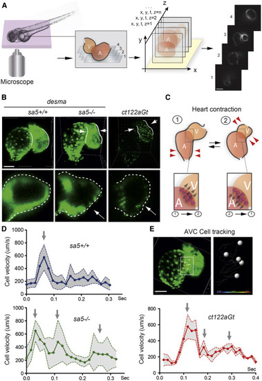
Heart Biomechanics Are Altered in the Absence of desma and in the Presence of Desmin Aggregates (A) 4D reconstructions of the heart-wall dynamics were obtained from 2D series recorded at a rate of 120 frames/s (fps) in successive planes. Periodic contractions were reconstructed in three dimensions using post acquisition synchronization of 48-hpf embryonic hearts. Scale bar, 30 μm. (B) Comparison of myocardial wall shape in desmasa5+/+, desmasa5−/−, and ct122aGt 4D reconstructed hearts shows a squeezing of the ventricle in both mutants ( Movies S2, S3, and S4). The lower panels show a zoom of an optical transverse section through the middle of the ventricle of desmasa5+/+, desmasa5−/−, and ct122aGt revealing the squeezing in absence of functional Desma (red arrows). Scale bar, 30 μm. (C) Schematic drawing recapitulating the myocardial movements associated with heart contraction and the subsequent movement of the atrioventricular canal (AVC). Examples of AVC cells tracks are shown in the lower panel. Their velocity was used as readout of the global contraction pattern of the heart. Scale bar, 30 μm. (D) Graph representing the velocity of myocardial cell motion in the AVC region following 3D cell tracking in desmasa5+/+, desmasa5−/−, and ct122aGt. (E) Individual nuclei were tracked automatically and their speed was extracted. ct122aGt indicates desmact122aGt homozygous embryos. The error bars correspond to the SEM. |
| Gene: | |
|---|---|
| Fish: | |
| Anatomical Term: | |
| Stage: | Long-pec |
| Fish: | |
|---|---|
| Observed In: | |
| Stage: | Long-pec |