
Fig. 7
- ID
- ZDB-FIG-170315-25
- Publication
- Ye et al., 2015 - Endoderm convergence controls subduction of the myocardial precursors during heart-tube formation
- Other Figures
- All Figure Page
- Back to All Figure Page
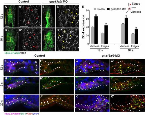
Epithelial organization of myocardial cells is disrupted in Gα13 morphants. (A-E) Whole-mount ZO-1 immunostaining was performed in embryos indicated at 12 s and 16 s. (A-D) Projections of confocal z-stacks of the right lateral mesoderm showing ZO-1 expression (gray) in the nkx2.5:Kaede-expressing (myocardial) cells (green). (A′-D′) Higher-magnification views of areas shown in boxes in A-D. The vertices were defined as regions where membranes from three adjacent cells come into contact, and the edges as the cell periphery excluding the vertices. Yellow dashed line, midline; circles, vertices; arrowheads, edges. (E) Intensity of ZO-1 expression in myocardial cells at the vertices and edges in control embryos (54 cells from 7 embryos at 12 s and 25 cells from 4 embryos at 16 s) and gna13a/b MO-injected embryos (51 cells from 8 embryos at 12 s and 20 cells from 4 embryos). *P<0.001 versus control. Data are mean±s.e.m. (F-K) Transverse vibratome sections immunostained for Kaede (magenta), ZO-1 (green), actin (Rhodamine-Phalloidin, red) and nuclei (DAPI, blue). Yellow dashed line indicates midline; white dashed lines outline myocardial cells; white and yellow arrowheads indicate normal and ectopic ZO-1 expression, respectively. Scale bars: 20 µm. |