
Fig. 4
- ID
- ZDB-FIG-170220-27
- Publication
- Brunt et al., 2016 - Building Finite Element Models to Investigate Zebrafish Jaw Biomechanics
- Other Figures
- All Figure Page
- Back to All Figure Page
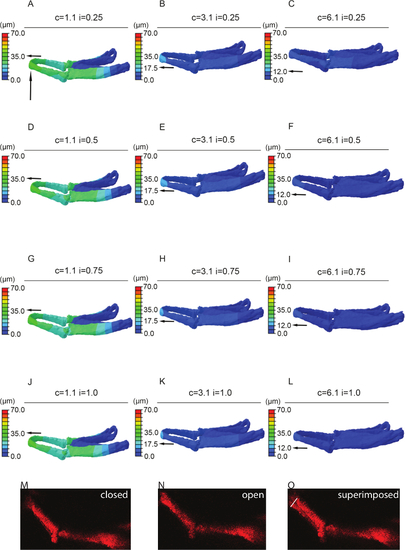
Sensitivity testing. FE-model simulating jaw displacement in 5dpf zebrafish for different cartilage and interzone Young's moduli. Jaw displacement (open to closed in µm) is marked on the jaw; recorded using the color key. Each model (A-L) has a different combination of cartilage (c = 1.1, 3.1, or 6.1 MPa) or interzone (i = of 0.25, 0.5, 0.75, or 1 MPa) properties. Horizontal black arrow highlights the value of jaw displacement at the tip of the Meckel's cartilage (represented by the vertical black arrow). M and N stills from videos of 5 dpf larvae showing minimum, i.e., jaw closed (M) and maximum, i.e., jaw fully open (N) with the two superimposed (O) - white line on O represents the displacement (of 43 µm). In this case relative cartilage properties of 1.1 with an interzone of 0.25 (A) best match the displacements seen in live fish (O). Panels A-L of this figure have been previously published in Brunt et al. 15. |