
Fig. S2
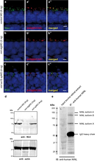
Specificity of the anti-Ninl antibody. (a) Indirect immunohistochemical staining using anti-Ninl antibody on 4 dpf retinal cryosections of control MO-injected larvae (green signal) shows punctate staining, partially overlapping with the ciliary marker anti-polyglutamylated tubulin (a', a”, red signal). (b) Indirect immunohistochemical staining using anti-Ninl antibody on 4 dpf retinal cryosections of ninl atgMO-injected larvae (b, green signal) along with the ciliary marker anti-polyglutamylated tubulin (b', b”, red signal). Specific Ninl-immunofluorescence is largely abolished in ninl morphants, whereas the polyglutamylated tubulin signal is still detected. (c) Indirect immunohistochemical staining of anti-Ninl on 4 dpf retinal cryosections of ninl ex15 spMO-injected larvae (green signal) along with the ciliary marker anti-polyglutamylated tubulin (c', c”, red signal). Specific Ninl-immunofluorescence was still detected but at a diminished level in ninl morphants whereas the polyglutamylated tubulin signal was unaltered. Nuclei are stained with DAPI (blue signal). Scale bars: 4 μm. (d) Western blot analysis using protein extracts obtained from 100 zebrafish larvae injected with either control MO (6ng), ninl atgMO (2ng) or ninl ex15 spMO (4ng). A specific product was detected with a molecular weight of ~80kDa in control MO-injected larvae (left panel). This band was almost completely abolished in the ninl atgMO-treated larvae, but was still detected in ninl spMO-injected larvae although with a slightly diminished intensity. Anti-actin antibodies were used as a loading control (right panel). (e) Immunoprecipitation from bovine retinal extracts with anti-human NINL antibody detects 3 bands, the strongest being of the same size as the band found on Western blot of zebrafish lysates (~80 kDa). |