
Fig. 3
- ID
- ZDB-FIG-170125-7
- Publication
- Cepero Malo et al., 2017 - The Zebrafish Anillin-eGFP Reporter Marks Late Dividing Retinal Precursors and Stem Cells Entering Neuronal Lineages
- Other Figures
- All Figure Page
- Back to All Figure Page
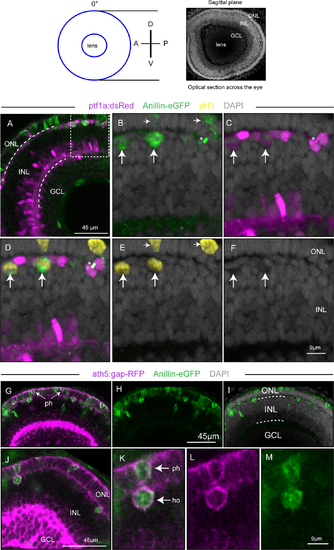
Visualisation of Anillin-eGFP labelled cycling cells in the post-embryonic central retina at 3 dpf. Top panel: view of the central retina in the sagittal plane (0°, eye facing the viewer). The blue circles represent an optical z-section across the eye corresponding to the blue line in the top panel of Fig 4. (A-F) Optical section from a z-stack through the retina of an 3 dpf, anillin:anillin-eGFP/ptf1a:dsRed transgenic zebrafish taken in the sagittal plane. (B-F) Magnification of the area delineated by the dotted rectangle in (A). The vertical arrows in (B-F) point at dividing horizontal cells that are (ptf1a)dsRed positive (magenta in C,D), Anillin-eGFP positive (green in B) and pH3 positive (yellow in D,E), and at their corresponding nuclei (F). The asterisk in (B-D) highlights an Anillin-eGFP labelled midbody between daughter cells in late cytokinesis, not depicted by pH3. The horizontal arrows in (B) and (E) point at two Anillin-eGFP positive (green in B) and pH3 positive (yellow in E) mitotic nuclei in the ONL. (G-I) and (J-M) represent two different optical sections from a z-stack through the retina of an 3 dpf, anillin:anillin-eGFP/atoh7:gap-RFP transgenic zebrafish in the sagittal plane. The arrows in (G) and (K) point at atoh7:gap-RFP expressing (magenta) photoreceptor and horizontal cell precursors that are also Anillin-eGFP (green) positive, indicating that they are in the cell cycle. GCL, ganglion cell layer; INL, inner nuclear layer; ONL, outer nuclear layer; ph, photoreceptor cell; ho, horizontal cell. |