
Fig. 4
- ID
- ZDB-FIG-170118-10
- Publication
- Pinzon-Olejua et al., 2017 - Cre inducible site-specific recombination in zebrafish oligodendrocytes
- Other Figures
- All Figure Page
- Back to All Figure Page
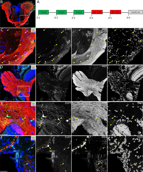
Conditional CreERT2-mediated recombination in Tg(mbpa:mCherry-T2A-CreERT2) in the adult Zebrafish CNS. A: Schematic representation of treatments applied to achieve conditional CreERT2-mediated recombination with the temperature-inducible, Cre-dependent reporter line Tg(hsp70l:loxP-DsRed-loxP-EGFP). B: Cross-section of the diencephalon with optic tracts and the rostral optic tectum immunostained for MBP (red) and EGFP (green). Scale bar, 100 µm. C: Higher magnification of the area depicted (white rectangle) in B showing presumptive oligodendrocytes (marked by arrowheads) after recombination expressing EGFP and co-stained with MBP (red). Scale bar, 50 µm. D: Cross-section of the retina with the exit point of the optic nerve immunostained for EGFP (green) and MBP (red), showing recombined cells (EGFP) within the MBP-expressing optic nerve. Scale bar, 50 µm. E: Higher magnification of the area depicted (white rectangle) in C showing EGFP-expressing cells (yellow arrowheads) co-expressing MBP. Scale bar, 25 µm. F: Cross-section of the dorsal telencephalon showing presumptive mature oligodendrocytes after recombination expressing EGFP (yellow arrowheads), co-immunostained with MBP (red) and located along the MBP-positive lateral olfactory tract (yellow dotted line) and the dorsal part of the entopeduncular nucleus. DAPI stains nuclei. Scale bar, 25 µm. |