
FIGURE
Fig. S1
- ID
- ZDB-FIG-161230-7
- Publication
- Nichols et al., 2016 - Ligament versus bone cell identity in the zebrafish hyoid skeleton is regulated by mef2ca
- Other Figures
- All Figure Page
- Back to All Figure Page
Fig. S1
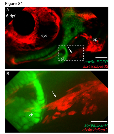
The anterior end of the operculohyoid ligament terminates at the ceratohyal cartilage. (A and B) 6 days post fertilization (dpf) sox9a:EGFP;alx4a:dsRed2 double transgenic larvae were live imaged by confocal microscopty. The dashed region in A is enlarged in B. The operculohyoid ligament is marked with an arrow, the ceratohyal (ch) cartilage, and the opercle (op) bone are indicated and the eye is shown for reference. Both images are projections of multiple confocal sections and lateral views where anterior is towards the left and dorsal is upward. Scale bars are 100µm in A, 25 µm in B. |
Expression Data
| Genes: | |
|---|---|
| Fish: | |
| Anatomical Terms: | |
| Stage: | Day 6 |
Expression Detail
Antibody Labeling
Phenotype Data
Phenotype Detail
Acknowledgments
This image is the copyrighted work of the attributed author or publisher, and
ZFIN has permission only to display this image to its users.
Additional permissions should be obtained from the applicable author or publisher of the image.
Full text @ Development