
Fig. 4
- ID
- ZDB-FIG-161222-11
- Publication
- Eum et al., 2016 - 3D Visualization of Developmental Toxicity of 2,4,6-Trinitrotoluene in Zebrafish Embryogenesis Using Light-Sheet Microscopy
- Other Figures
- All Figure Page
- Back to All Figure Page
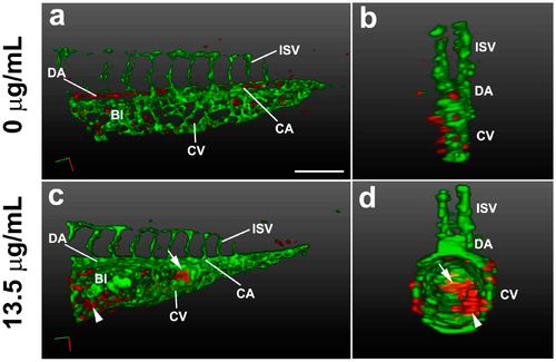
3D light-sheet/SPIM imaging of the TNT circulation defect. The vasculature (green) and blood cells (red) of the posterior trunk were visualized in Tg(gata1:DsRed/fli1a:EGFP) embryos at 38 hpf. 3D reconstructions are shown at lateral view (a,c) and transverse view (b,d), using Arivis software (surface function). (a,b) Embryos treated for 33 h with control E3 media; (c,d) Embryos treated for 33 h with pink water containing 13.5 µg/mL TNT showing swollen body structure with abnormal blood islands (arrowhead) and blood accumulation (arrow). 20X water lens, 0.8 zoom. Scale bar, 150 µm. BI, blood Island; CA, caudal artery; CV, caudal vein; DA, dorsal aorta; ISV, intersegmental vessel. The 3D movies of the reconstructions are presented in Supplementary video 2A,B. |