
Fig. 2
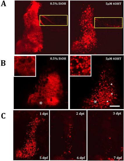
Tamoxifen-induced PC death in PC-ATTACTM fish. (A,B) Incubation of 4 dpf PC-ATTACTM embryos (n=6) in 4OHT (5×10⁻⁶M) for 16 h induces cell death of PCs in larvae at 5 dpf compared with controls treated with 0.5% EtOH. Cell ablation was visible by way of large amounts of PC debris (white arrowheads in B). The extensive dendritic layer was completely disrupted (the asterisks mark the region shown in the inset in B) and the projections of the cerebello-octavolateralis were fragmented (yellow square in A). (C) PC-ablated larvae re-analyzed 24, 48 and 72 h after tamoxifen treatment show decreasing amounts of red fluorescent PC debris and a lack of fast PC recovery. Scale bars: 20 µm; dpt, days post-treatment. |