
Fig. 7
- ID
- ZDB-FIG-161202-29
- Publication
- Jacob et al., 2016 - Evolution and Expression of Paxillin Genes in Teleost Fish
- Other Figures
- All Figure Page
- Back to All Figure Page
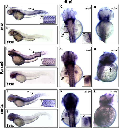
Expression profiles for Paxillin orthologs in the zebrafish embryo two days post-fertilization. (A-D) At 48 hpf, a pxna antisense probe detected specific expression in myotomes (m) (A) and the pectoral fin (pf) bud (C; dorsal view), when compared to background staining in the head revealed by control pxna (B), pxnb (F) and pxnb-ins (J) sense probes. A' and C' depict zoomed-in boxed regions in A and C. No pxna expression was observed in the heart (D; ventral view). (E-H) A pan-pxnb probe detected expression in vertical myosepta (vm) (E), pectoral fin bud (G) and heart (h) (H). E' and G' depict zoomed-in boxed regions of E and G. (I-L) A pxnb-ins-specific antisense probe detected expression in vertical myosepta (I) and pectoral fin bud (K), but no expression in the heart (L). I' and K' depict zoomed-in boxed region of I and K. |
| Genes: | |
|---|---|
| Fish: | |
| Anatomical Terms: | |
| Stage: | Long-pec |