
Fig. 2
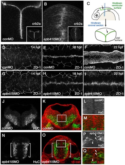
Epb41l5 is required for the formation of the hindbrain ventricle and the proper localization of neurons. (A-C) Coronal sections showing mislocalization of Crb2a in the hindbrain of epb41l5 morphants. Whereas Crb2a is restricted to a well-defined apical/ventricular surface in wild-type embryos injected with control morpholino (MO) (conMO), Crb2a is diffusely expressed in epb41l5 morphants (epb41l5MO). Lower-magnification images are shown in insets. Diagram in C illustrates approximate positions of coronal sections in the hindbrain. (D-I) Failure of organization of ZO-1-positive AJs and formation of the hindbrain ventricle in epb41l5 morphants. Confocal images were taken from the dorsal side of embryos. The anterior is to the left and the posterior is to the right. In control MO embryos, accumulation of ZO-1+ puncta at 14hpf is gradually aligned to the midline of the hindbrain and forms a continuous line at 18hpf. The hindbrain ventricle inflates at 22hpf. In epb41l5 morphants, ZO-1 accumulation at 14hpf is gradually aligned to the midline of the hindbrain but fails to form a continuous line at 18hpf. The hindbrain ventricle is not formed at 22hpf. (J-Q) Coronal sections showing mislocalization of HuC-expressing neurons in epb41l5 morphants. In control MO embryos, HuC-positive neurons are localized at the pial region of the hindbrain and do not have contacts with the ventricular surface. In epb41l5 morphants, a fraction of HuC-positive cells were located near the midline of the hindbrain and maintain contacts with the prospective ‘ventricular’ surface. Boxed areas in K and O are shown at higher magnification in L,M and P,Q, respectively. Brackets indicate HuC-positive neurons which are still localized near the basal/pial surface of the hindbrain. Arrowheads indicate HuC-positive cells which are localized near the midline. |