
Fig. 3
- ID
- ZDB-FIG-160927-49
- Publication
- Zhao et al., 2016 - Enhanced angiogenesis, hypoxia and neutrophil recruitment during Myc-induced liver tumorigenesis in zebrafish
- Other Figures
- All Figure Page
- Back to All Figure Page
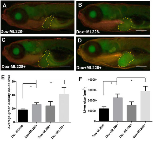
Stimulation of tumorigenic liver growth by hypoxia. TO(Myc) and Tg(phd3::EGFP) double transgenic larvae were generated and induced by Dox for 4 days from 4 dpf to 7 dpf. (A-D) Images of liver hypoxia as indicated by EGFP expression in dashline-circled liver areas. A non-Dox treated control is shown in (A) and a Dox treated larva is in (B). 0.5 µM ML228 was used to enhance hypoxia and representative images are shown in (C,D) in the absence or presence of Dox, respectively. The original magnification was 20x. (E) Quantification of level of hypoxia as indicated by average GFP green density inside the liver. (F) Quantification of 2D liver size. The quantitative data were based on 5 samples per concentration group. Data are represented as mean ± SD. Astrisks indicate significant difference with P-value < 0.05 by unpaired t-test statistical analysis. Scale bars = 100 µm. |
| Gene: | |
|---|---|
| Fish: | |
| Conditions: | |
| Anatomical Term: | |
| Stage: | Days 7-13 |
| Fish: | |
|---|---|
| Condition: | |
| Observed In: | |
| Stage: | Days 7-13 |