
Fig. 1
- ID
- ZDB-FIG-160713-6
- Publication
- Meyer et al., 2016 - Microarray Noninvasive Neuronal Seizure Recordings from Intact Larval Zebrafish
- Other Figures
- All Figure Page
- Back to All Figure Page
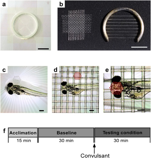
Tools and steps for larva-mounting and timeline of recording. a: Electrode chamber used for recordings (scale: 10 mm). b: Nylon mesh and slice anchor used to hold larva in place (scale: 5 mm). c: Placement of larva onto its dorsal side into the chamber with a drop of water (scale: 300 µm). d: Securing larva with nylon mesh (scale: 300 µm); microelectrode array is highlighted with red dots for enhanced visibility in this photo and the next. e: Larva positioned with head onto the microelectrode array (scale 300 µm). f: Timeline for experiments-each starting with a 15 min acclimation period, a 30 min control recording, and a 30 min recording time after drug application. |