
Fig. 3
- ID
- ZDB-FIG-160706-24
- Publication
- Feng et al., 2016 - HIF-1α and HIF-2α induced angiogenesis in gastrointestinal vascular malformation and reversed by thalidomide
- Other Figures
- All Figure Page
- Back to All Figure Page
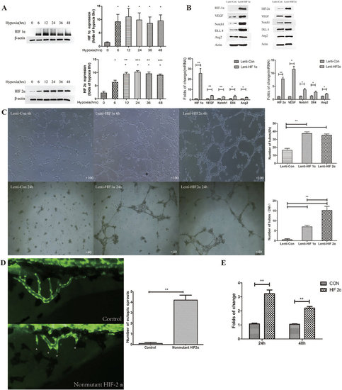
(A) Western blot determinations of HIF-1α and HIF-2α expression at different time points of hypoxia. *P < 0.05, **P < 0.01 vs. 0 hour. (B) The effect of HIF-1α and HIF-2α overexpression on the expression of VEGF, Notch1, DLL4, and Ang2. Western blot and RT-PCR demonstrated that HIF-1α and HIF-2α overexpression increased the expression of VEGF, Notch1, DLL4, and Ang2 protein and mRNA. *P < 0.05, **P < 0.01 vs. control. (C) Influence of HIF-1α and HIF-2α overexpression on angiogenesis 6 and 24 h after transfection of Lenti-HIF-1α and Lenti-HIF2α. Tube formation was enhanced 6 and 24 h after transfection. **P < 0.01 vs. control. (D) Fluorescence microscope observations of subintestinal vein sprouting in normal and HIF-2α-overexpressing zebrafish. *Indicates subintestinal vascular sprouts. HIF-2α overexpression significantly increased the number of subintestinal vascular sprouts. **P < 0.01 vs. control plasmid. (E) Dual luciferase reporter gene assay demonstrated that HIF-2α enhanced VEGF promoter activity. **P < 0.01 vs. control plasmid. |