
Fig. S1
- ID
- ZDB-FIG-160603-9
- Publication
- Foglia et al., 2016 - Multicolor mapping of the cardiomyocyte proliferation dynamics that construct the atrium
- Other Figures
- All Figure Page
- Back to All Figure Page
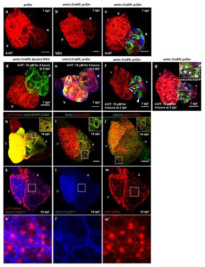
amhc:CreER-mediated recombination of the priZm cassette fluorescently labels individual zebrafish atrial cardiomyocytes. (a-c) Surface myocardium images of 7 dpf priZm hearts with and without the amhc:CreER transgene and after 4-HT or vehicle (VEH) treatment. (d) Surface myocardium of a 7 dpf amhc:CreER; βactin2:RSG heart after treatment with 4-HT at 3 dpf. (e) Surface myocardium of a 7 dpf cmlc2:CreER; priZm heart after treatment with 4-HT at 3 dpf. Scale bars are 50 µm. (f) Surface myocardium of a 7 dpf amhc:CreER; priZm heart from a larva treated with 4-HT at 3 dpf. Labeled cardiomyocytes are present in singlet (arrowhead) and doublet (arrows) clones. (g) Atrial cardiomyocytes from a 7 dpf amhc:CreER; priZm heart with nuclei marked by cmlc2:H2-GFP. Scale bars are 50 µm. (h) 3D reconstruction of a 14 dpf heart from an animal expressing the cmlc2:dsRed and cmlc2:EGFP-CAAX transgenes. Non-myocardial regions of the atrial wall are indicated (dotted lines). (i, j) Surface myocardium of 14 dpf hearts expressing βactin2:RSG and immunostained for Atrial myosin heavy chain (Amhc) or Laminin, respectively. A non-myocardial region of the atrial wall is indicated with dotted lines. Laminin immunofluorescence covers the entire atrial surface. Scale bars are 50 µm. (k-m) Surface of a 14 dpf heart from an animal expressing the epicardial reporter tcf21:dsRed and the myocardial reporter βactin2:tagBFP. Epicardial cells coat the entire atrial surface, whereas there are myocardial gaps. White boxes indicate magnified area. |