
Fig. 6
- ID
- ZDB-FIG-160512-31
- Publication
- Stil et al., 2016 - Neuronal labeling patterns in the spinal cord of adult transgenic Zebrafish
- Other Figures
- All Figure Page
- Back to All Figure Page
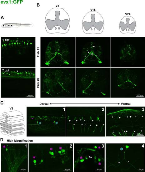
Labeling pattern of GFP in the spinal cord of adult evx1:GFP transgenic fish. In evx1:GFP embryos, GFP is driven in commissural interneurons (asterisk, A). In adults, cell bodies that project ventrally to the contralateral side along commissural paths are found adjacent to the central canal (asterisk, B). Some GFP-positive fibers sparsely covered sections (arrows, B, C). Some big bundles start above the central canal and go down to the ipsilateral ventral root (arrowheads, B, C), and their cell bodies are found around the central canal (magenta asterisks; C; D). Higher magnification shows other cells (blue asterisk, D) with processes extending to both rostral and caudal directions (arrowheads, D). |