
Fig. 5
- ID
- ZDB-FIG-160418-5
- Publication
- Diaz-Tellez et al., 2016 - The Zebrafish scarb2a Insertional Mutant Reveals a Novel Function for the Scarb2/Limp2 Receptor in Notochord Development
- Other Figures
- All Figure Page
- Back to All Figure Page
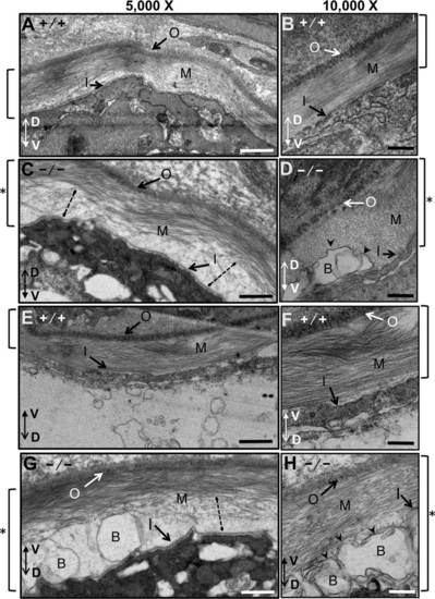
Notochordal basement membrane in WT and mutant zebrafish embryos at 3 dpf analyzed by electron microscopy. All images are transverse cross-sections from near somite 16. For this figure, images were oriented so the outer layer of the basal membrane is at the top, and the dorso-ventral axis of the larvae is indicated by a double arrow. A-D: Views of the basal membrane on the dorsal side of the notochord. E-H: Views of the basal membrane on the ventral side of the notochord. A,B,E,F: Wild-type embryos. C,D,G,H: scarb2ahi1463Tg mutant zebrafish embryos at 3 dpf. Images were obtained at either 5,000X or 10,000X. Square brackets to the sides of each figure indicate the thickness of the basement membrane, which was thicker in the mutants (see asterisks). Dotted lines in sections from mutant embryos show regions where collagen appears to be not properly assembled. Blisters (arrowheads) appear in mutant basal membranes just above the basal lamina of the inner layer. Outer basal membrane (O), medial basal membrane (M), inner basal membrane (I) and blisters (B). Scale bars = 500 nm. |