
FIGURE
Fig. S1
- ID
- ZDB-FIG-160405-7
- Publication
- Webb et al., 2016 - Persistence, period and precision of autonomous cellular oscillators from the zebrafish segmentation clock
- Other Figures
- All Figure Page
- Back to All Figure Page
Fig. S1
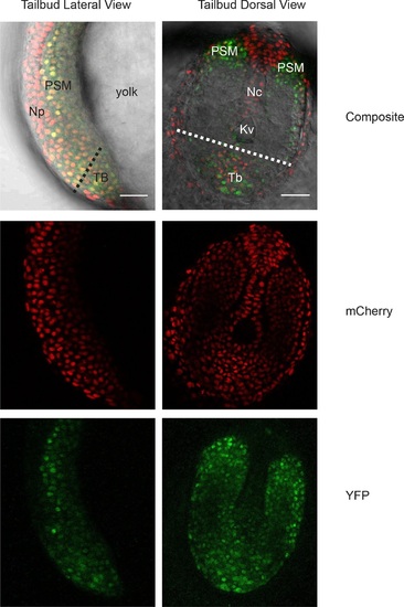
Her1-YFP-expressing cells in the zebrafish tailbud.A confocal section through the tailbud of a Her1-YFP and Histone 2A-mCherry expressing 8-somite stage Looping zebrafish embryo in both lateral and dorsal orientations. The approximate location of the segmentation clock cells removed by surgery to generate the tailbud explants and single cell cultures is shown with the dashed line. Nuclei are shown in red and YFP in green. Scale bar = 50 µm. Kupffer’s vesicle (Kv), notochord (Nc), presomitic mesoderm (PSM), tailbud (Tb), neural progenitors (Np), yolk cell (yolk). |
Expression Data
Expression Detail
Antibody Labeling
Phenotype Data
Phenotype Detail
Acknowledgments
This image is the copyrighted work of the attributed author or publisher, and
ZFIN has permission only to display this image to its users.
Additional permissions should be obtained from the applicable author or publisher of the image.
Full text @ Elife