
Fig. 1
- ID
- ZDB-FIG-160401-7
- Publication
- Ott et al., 2016 - Pronephric tubule morphogenesis in zebrafish depends on Mnx mediated repression of irx1b within the intermediate mesoderm
- Other Figures
- All Figure Page
- Back to All Figure Page
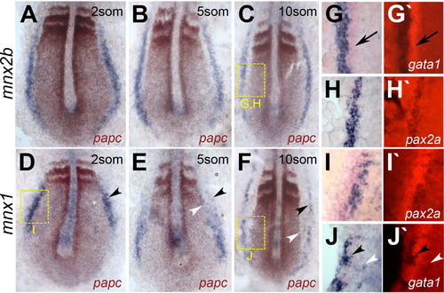
Distinct expression patterns for mnx2b and mnx1 in intermediate mesoderm. (A-F) Flat-mounts of embryos co-stained for mnx genes (blue) and papc (brown). Expression of mnx2b (A-C) and mnx1 (d-F) in the IM is present in 2 somite (A, D), 5 somite (B, E) and 10 somite stage (C,F) embryos. Black arrows point to the gata1 signal, black arrowheads indicate the lateral edge of mnx staining, white arrowheads the medial edge of mnx1 staining. Note that anterior mnx1 signals at 5 and 10 somite stage forms a lateral (black arrowhead) and a medial stripe (white arrowhead). Yellow boxes indicate positions of higher resolution images shown in G-J. (G-J) Double-ISH stainings for mnx genes (blue) with intermediate mesoderm markers (red) for nephrogenic (pax2.1; G-I) and hematopoietic cell lineages (gata1; J, J′). Shown are brightfield (G, H) and fluorescence images (G′-J′). Mnx2b expression is excluded from gata1 labeled hematopoietic regions (arrow in G, G′), but completely overlaps with the pronephric marker pax2.1 (H, H′). In contrast, mnx1 signals are flanking or intermingled with signals for pax2.1 (2 somite stage; I, I′) and gata1 (10 somite stage, J, J′). |
Reprinted from Developmental Biology, 411(1), Ott, E., Wendik, B., Srivastava, M., Pacho, F., Töchterle, S., Salvenmoser, W., Meyer, D., Pronephric tubule morphogenesis in zebrafish depends on Mnx mediated repression of irx1b within the intermediate mesoderm, 101-14, Copyright (2016) with permission from Elsevier. Full text @ Dev. Biol.