
Fig. S4
- ID
- ZDB-FIG-160224-10
- Publication
- Ulrich et al., 2016 - Reck enables cerebrovascular development by promoting canonical Wnt signaling
- Other Figures
- All Figure Page
- Back to All Figure Page
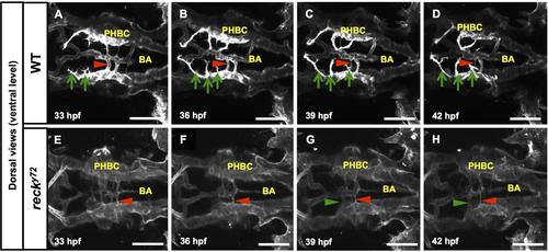
In recky72 the PHBCs are deficient in CtA sprout formation and display abnormal avc dynamics. Time-lapse stills (dorsal views, ventral-level) of Hb vascular development (visualized with Tg(kdrl:GFP)1a116) in WT (A-D) and recky72 (E-H) from 33 to 42 hpf. Anterior, left; right side, up. Green arrows, developing CtA sprouts on the embryo’s left side. Green arrows, de novo avc formation. Red arrowheads, regressing avc. In this WT (A-D) three filopodia-rich CtA sprouts launch from the dorsal side of the left PHBC, one PHBC-BA avc thins and regresses but no new avc develop. In this recky72 (E-H) filopodial activity is absent from the dorsal face of the PHBCs and, accordingly, no CtA-like, dorsally projecting sprouts emerge. On the mutant’s left side a PHBC-BA avc regresses and another forms. There is no evidence that the mutant’s supernumerary avc are mistargeted CtAs unable to invade the brain. Scale bars: 100 µm. See Movies S2A-S2B. |