
Fig. S4
- ID
- ZDB-FIG-160205-59
- Publication
- Nikolaou et al., 2015 - Lamination Speeds the Functional Development of Visual Circuits
- Other Figures
- All Figure Page
- Back to All Figure Page
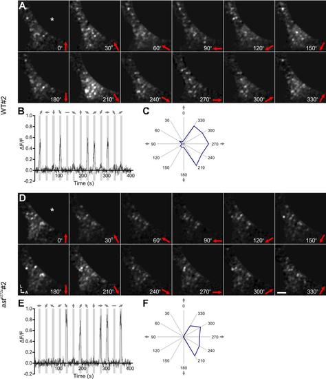
DS-RGC axons are diffusely distributed in the tectal neuropil of individual astray mutants (related to Figure 2). (A-D) Functional maps generated from single Tg(Isl2b:Gal4;UAS:SyGCaMP3) larvae showing the spatial distribution of DS-RGC subtypes in the tectal neuropil of WT larvae at 4 dpf (A) and 7 dpf (B), and astti272z larvae at 4 dpf (C) and 7 dpf (D). Four examples of WT and astti272z larvae are shown, respectively. Voxel brightness is proportional to the summed incidence of each functional subtype across all experiments 3 performed in each individual fish. Dashed lines indicate the position of the skin overlaying the tectum. Scale bar represents 20 µm. A, anterior; L, lateral. |