
Fig. 1
- ID
- ZDB-FIG-151228-11
- Publication
- Mendieta-Serrano et al., 2015 - Spatial and temporal expression of zebrafish glutathione peroxidase 4 a and b genes during early embryo development
- Other Figures
- All Figure Page
- Back to All Figure Page
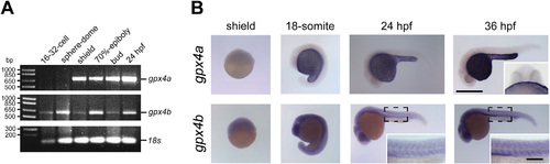
Expression patterns of gpx4a and gpx4b during early zebrafish embryo development. A, RT-PCR expression analysis of gpx4a, gpx4b and 18s in zebrafish embryos at different developmental stages, 16- to 32-cell stage, sphere to dome, shield, 70%-epiboly, bud and 24 hpf. B, in situ hybridization analysis of gpx4a and gpx4b during early zebrafish embryo development. Upper inset, 36 hpf embryos were mounted in agarose and manually sliced to show the signal from the in situ localization of gpx4a is found in the periderm covering the yolk cell. Lower insets, highlight the gpx4b localization in the myotomes. Scale bar 1 mm for all stages and 250 µm for insets. |
| Genes: | |
|---|---|
| Fish: | |
| Anatomical Terms: | |
| Stage Range: | 16-cell to Prim-25 |
Reprinted from Gene expression patterns : GEP, 19(1-2), Mendieta-Serrano, M.A., Schnabel-Peraza, D., Lomelí, H., Salas-Vidal, E., Spatial and temporal expression of zebrafish glutathione peroxidase 4 a and b genes during early embryo development, 98-107, Copyright (2015) with permission from Elsevier. Full text @ Gene Expr. Patterns