
Fig. 2
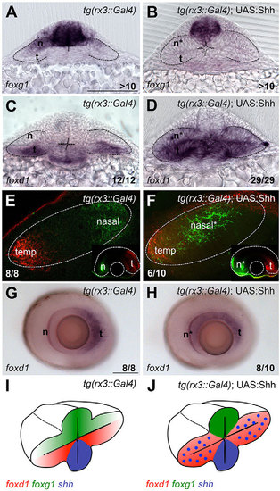
Ectopic Hh activity in the optic vesicle promotes temporal fate. (A-D,G,H) Expression of foxg1 (A,B) and foxd1 (C,D,G,H) in the genotypes specified in the panels. (E,F) Retinotectal projections traced with DiI/DiO to label nasal (n; green) and temporal (t; red) projections. Main panels show the tectum with anterior to the left; insets show the corresponding eye. (I,J) Schematic of the phenotypic outcome of foxg1/foxd1 expression in the conditions shown in the figure. A-D are frontal views; G and H are lateral views of dissected eyes. All embryos are at 10-12ss except those in E-H, which are 6dpf. Scale bars: 100µm. Numbers in the bottom-right of each panel indicate the number of embryos with the phenotype shown out of the total number of embryos analysed. n*, defective nasal domain. Dashed lines outline the optic vesicles (A-D), the tectum (E,F) and the optic cup (insets in E,F). |