
Fig. 6
- ID
- ZDB-FIG-151119-34
- Publication
- Walton et al., 2015 - The Macrophage-Specific Promoter mfap4 Allows Live, Long-Term Analysis of Macrophage Behavior during Mycobacterial Infection in Zebrafish
- Other Figures
- All Figure Page
- Back to All Figure Page
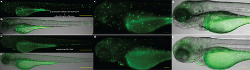
mfap4:iCre:p2A-tdTomato mediates macrophage-specific gene expression of loxP-containing transgenes. a-d. Zebrafish larva at 3 dpf expressing both Tg(β-actin2:loxP-DsRed-STOP-loxP-EGFP)s928 and Tg(mfap4:iCre:p2A-tdTomato)xt8; a,b. 50x magnification of GFP fluorescence and merge with brightfield, respectively. c,d. 200x magnification of the head, yolk sac, and anterior portion of the gut. e-h. Zebrafish larva at 3 dpf expressing Tg(mfap4:dLanYFP-CAAX)xt11 for comparison. e,f. 50x magnification of dLanYFP fluorescence and merge with brightfield, respectively. g,h. 200x magnification of the head, yolk sac, and anterior portion of the gut. Note that in both larvae, fluorescent protein expression is restricted to macrophages all along the body, with a particular enrichment in the head. Both larvae also exhibit expression within the caudal hematopoietic tissue (CHT). The larvae in a-d show a reduced number of fluorescent cells in the nascent macrophage population that predominates in the CHT region, and modestly reduced fluorescent cell population number in regions where mature macrophages have migrated throughout the body. Scale bars for 50x images = 500 µm; scale bars for 200x images = 100 µm. |