
Fig. S3
- ID
- ZDB-FIG-151102-6
- Publication
- Blum et al., 2015 - Retinoic acid signaling spatially restricts osteoblasts and controls ray-interray organization during zebrafish fin regeneration
- Other Figures
- All Figure Page
- Back to All Figure Page
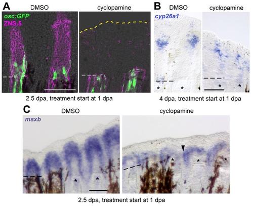
Absence of preosteoblasts causes blastema expansion. Cyclopamine treatment starting at 1 dpa results in preosteoblast-free blastema and lateral expansion of blastema cells, but normal cyp26a1 expression. (A) IHC for ZNS-5 and GFP in osc:gfp fish demonstrates lack of GFP+ preosteoblasts in the blastema of cyclopamine treated fish at 2.5 dpa. Yellow dashed line indicates the distal blastema edge in cyclopamine treated fish. (B) WISH for cyp26a1 at 4 dpa reveals unaltered cyp26a1 expression in cyclopamine treated fish (C) WISH for msxb demonstrates lateral expansion of blastema cells into the interray tissue (arrowhead) in cyclopamine treated fish. Dashed lines indicate amputation plane. Scale bars: 100 µm. |