
Fig. 5
- ID
- ZDB-FIG-151030-32
- Publication
- Blum et al., 2015 - Osteoblast de- and redifferentiation is controlled by a dynamic response to retinoic acid during zebrafish fin regeneration
- Other Figures
- All Figure Page
- Back to All Figure Page
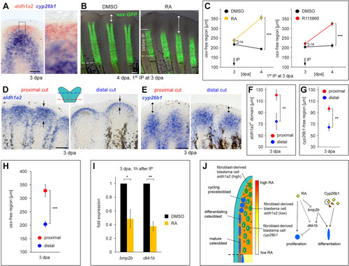
RA signaling keeps preosteoblasts in an undifferentiated state. (A) Distal blastema cells express aldh1a2 but not cyp26b1. Double WISH at 3dpa. (B,C) RA and R115866 injections at 3dpa block preosteoblast differentiation. (B) Live images of osx:gfp fish at 4dpa demonstrate an expanded distal osx-free region (doubled-headed arrow) one day after RA injection. (C) Length of the osx-free domain. (D-H) aldh1a2 expression (D,F), the distal cyp26b1-free domain (E,G) and the osx-free domain (H) extend further proximally in regenerates that had been amputated at a more proximal level. (D) WISH for aldh1a2 at 3dpa. Arrows indicate expression boundaries. (E) WISH for cyp26b1. Double-headed arrows indicate length of the cyp26b1-free region. (F-H) Length of the expression domain or of the distal expression-free region. (I) Injection of RA at 3dpa downregulates bmp2b and dkk1b expression (qPCR analysis). (J) Model for regulation of preosteoblast differentiation by RA signaling. Data are represented as mean±s.e.m. *P<0.05, **P<0.01, ***P<0.001. Dashed lines indicate amputation planes. Scale bars: 100µm. h, hours. |