
Fig. S4
- ID
- ZDB-FIG-151012-5
- Publication
- Swinburne et al., 2015 - Improved Long-Term Imaging of Embryos with Genetically Encoded α-Bungarotoxin
- Other Figures
- All Figure Page
- Back to All Figure Page
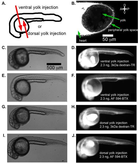
α-bungarotoxin protein injected into the yolk is distributed throughout the embryo. (A) Two injection strategies explored for α-bungarotoxin protein injection: either into the ventral side of the yolk or the dorsal side of the yolk of zebrafish embryos at 24 hpf. (B) Fluorescence from 2.3 ng Alexa-Fluor 594 conjugated α-bungarotoxin injected into the ventral yolk imaged by laser-scanning confocal microscopy. The peripheral yolk space appears continuous with the fluid entering the heart. DIC images (left) and fluorescent images (right) of representative embryos receiving ventral yolk injections of 2.3 ng 3 kDa dextran-Texas red (C-D), ventral yolk injections of 2.3 ng Alexa-Fluor 594 conjugated α-bungarotoxin (E-F), dorsal yolk injections of 2.3 ng 3 kDa dextran-Texas red (G-H), and dorsal yolk injections of 2.3 ng Alexa-Fluor 594 conjugated α-bungarotoxin (I-J). Scale bar in (C) applies for (C-J). |