
Fig. S1
- ID
- ZDB-FIG-151005-15
- Publication
- Li et al., 2015 - A transgenic zebrafish model for monitoring xbp1 splicing and endoplasmic reticulum stress in vivo
- Other Figures
- All Figure Page
- Back to All Figure Page
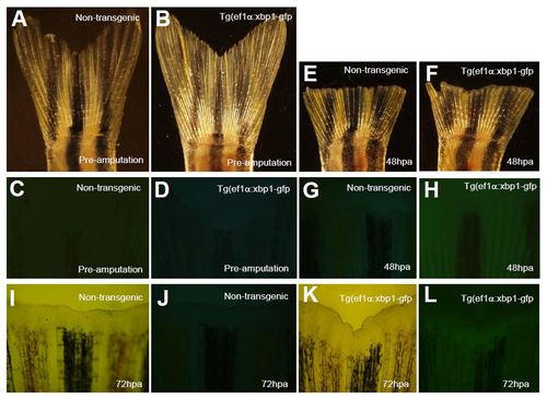
Analysis of Tg(ef1α:xbp1δ-gfp) expression during caudal fin regeneration. Non-transgenic and Tg(ef1α:xbp1δ-gfp) transgenic fish (4 months) were subjected to caudal fin amputation. XBP1Δ-GFP expression were analyzed at preamputation and 48 and 72 hours post amputation (hpa). A–D. Bright field (A, B) and whole mount fluorescent (C, D) images of caudal fins of non-transgenic and Tg(ef1α:xbp1δ-gfp) transgenic fish at preamputation. E–H. Bright field (E, F) and whole mount fluorescent (G, H) images of caudal fins of non-transgenic and Tg(ef1α:xbp1δ-gfp) transgenic fish at 48 hpa. I–L. Bright field (I, K) and whole mount fluorescent (J, L) images of caudal fins of non-transgenic and Tg(ef1α:xbp1δ-gfp) transgenic fish at 72 hpa. |
Reprinted from Mechanisms of Development, 137, Li, J., Chen, Z., Colorni, A., Ucko, M., Fang, S., Du, S.J., A transgenic zebrafish model for monitoring xbp1 splicing and endoplasmic reticulum stress in vivo, 33-44, Copyright (2015) with permission from Elsevier. Full text @ Mech. Dev.