
Fig. 3
- ID
- ZDB-FIG-151002-32
- Publication
- Cavanaugh et al., 2015 - Two developmentally distinct populations of neural crest cells contribute to the zebrafish heart
- Other Figures
- All Figure Page
- Back to All Figure Page
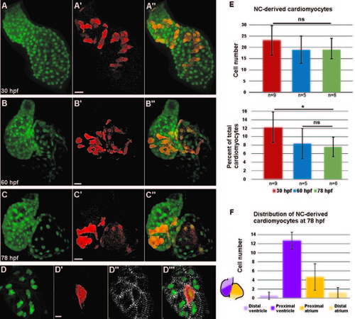
Distribution of neural crest-derived cardiomyocytes in developing zebrafish heart. (A–C′′) Confocal projections of NC:NfsB-mCherry; myl7:nucGFP embryonic hearts at 30 hpf (A–A′′), 60 hpf (B–B′′), and 78 hpf (C–C′′). (A–A′′) NC-derived mCherry positive cells are distributed along the primitive heart tube at 30 hpf. (B–C′′) As development progresses, mCherry+ cells become excluded from the poles of the heart. Scale bar=20 µm. (D–D′′′) Confocal image of the ventricle of a NC:NfsB-mCherry; myl7:nucGFP embryo at 72 hpf. Immunostaining for α-actinin (white) demonstrates well-developed sarcomeres in NC-derived cardiomyocytes. Scale bar=5 µm. (E) Quantification of the number (top) and the percentage (lower panel) of NC-derived cardiomyocytes (positive for both mCherry and nucGFP) at 30 hpf, 60 hpf and 78 hpf. (F) At 78 hpf NC-derived cardiomyocytes contribute primarily to the proximal ventricle (12.7±1.9) and proximal atrium (4.7±2.9), with very little contribution to the distal ventricle (0.5±0.8) and distal atrium (1.2±1.5) (n=6). *p<0.05; ns, not significant. |
Reprinted from Developmental Biology, 404(2), Cavanaugh, A.M., Huang, J., Chen, J.N., Two developmentally distinct populations of neural crest cells contribute to the zebrafish heart, 103-12, Copyright (2015) with permission from Elsevier. Full text @ Dev. Biol.