
FIGURE
Fig. 7
- ID
- ZDB-FIG-150928-21
- Publication
- McGorty et al., 2015 - Open-top selective plane illumination microscope for conventionally mounted specimens
- Other Figures
- All Figure Page
- Back to All Figure Page
Fig. 7
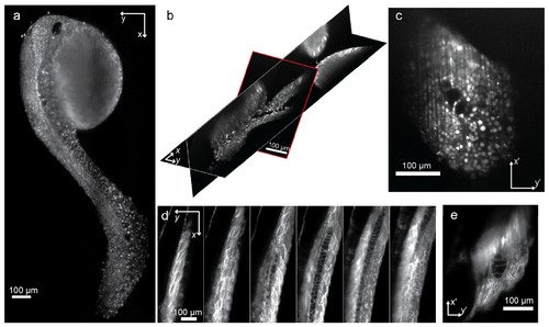
Imaging zebrafish in 96-well plates (a) One x-y slice of a 48 hpf zebrafish in a 96-well plate. All cell nuclei are labeled with mRFP. (b) Multiple slices from the same embryo shown in (a). The slice outlined in red is the raw camera image before any transformations. The other two slices show an x-y and x-z cut. (c) An image of the same embryo as recorded on the camera. (d) Six x-y slices through the tail of a zebrafish with tdTomato labeling of the cell membranes. Each slice is spaced 23 µm apart in z. (e) An image as recorded by the camera of the same embryo as shown in (d) (see Media 5). |
Expression Data
| Genes: | |
|---|---|
| Fish: | |
| Anatomical Terms: | |
| Stage: | Long-pec |
Expression Detail
Antibody Labeling
Phenotype Data
Phenotype Detail
Acknowledgments
This image is the copyrighted work of the attributed author or publisher, and
ZFIN has permission only to display this image to its users.
Additional permissions should be obtained from the applicable author or publisher of the image.
Full text @ Opt. Express