
Fig. 2
- ID
- ZDB-FIG-150928-1
- Publication
- Chen et al., 2015 - Invasiveness and metastasis of retinoblastoma in an orthotopic zebrafish tumor model
- Other Figures
- All Figure Page
- Back to All Figure Page
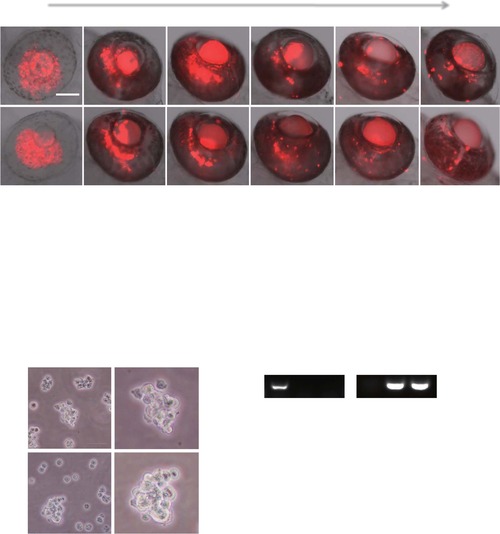
Formation of primary retinoblastoma in the zebrafish retina, and re-culture of implanted retinoblastoma cells from zebrafish in vitro. (a) Approximately, 150 DiI-labeled human (RB355) or mouse (SJmRBL-8) retinoblastoma cells (red color) were intravitreally implanted into each eye of zebrafish and the formation of primary tumors were kinetically monitored under fluorescent microscopy at different time points after tumor cell implantation. Bar = 100 µm. (b) Primary tumor areas were quantified using a digital Photoshop program. Approximately 30–40 tumor samples were used for quantification. (c) Transplanted SJmRBL-8 retinoblastoma cells are successfully isolated and cultured in vitro. The photographs at day 14 from inverted microscope showed the transplanted cells maintained their morphology and viability after injection. (d) Total RNA was extracted from zebrafish embryos, mouse SJmRBL-8 cells and the re-cultured cells, and RT-PCR was then used to analyze zebrafish and mouse GAPDH expression. (e) The expression of zebrafish and mouse GAPDH was determined by quantitative real-time PCR. |