
Fig. S1
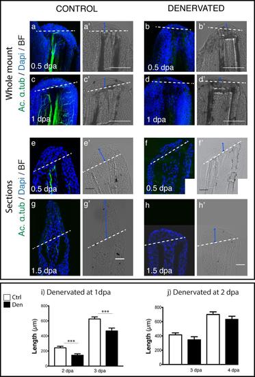
Analysis of the regenerative process of denervated pectoral fins. a-d) Staining for ac. α-tub and DAPI in whole mount fins. Staining with DAPI confirms that fins without innervation (b,d) have a WE with less epidermal cell layers than controls (a,c). e-h) Staining for ac. α-tub and DAPI in sections. Staining with DAPI shows that fins without innervation (e) have a WE with less epidermal cell layers than controls (f). After 1.5 dpa denervated fins (h) are not able to regenerate as the controls (g). a-h) Dashed lines mark amputation plane. i,j) Quantification of the length of regenerated tissue in fins denervated after amputation. Measurements of the length of regenerated tissue, taken from the amputation site to the most distal tip. i) There is a consistent significant reduction (***p < 0.0001) in the length of fins denervated at 1 dpa in relation to controls, both at 2 and 3 dpa. j) There is no significant reduction in the length of regenerates in fins denervated at 2dpa in relation to controls (fix at 3 dpa p = 0.33; fix at 4 dpa p = 0.07). |