
Fig. 3
- ID
- ZDB-FIG-150810-22
- Publication
- Briona et al., 2015 - Wnt/ß-catenin signaling Is required for radial glial neurogenesis following spinal cord injury
- Other Figures
- All Figure Page
- Back to All Figure Page
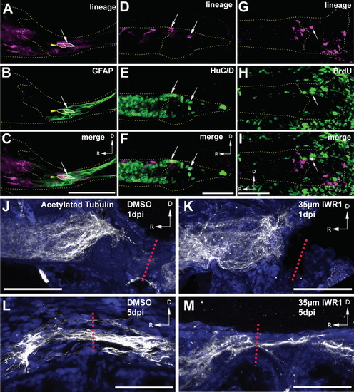
Representative images of gfap:CreERT2-labeled progeny in the regenerating blastema. (A–C) At 5 dpi one mCherry+ cell is GFAP+ (arrow), while a neighboring cell is GFAP (arrowhead). (D–F) At 5 dpi, multiple HuC/D+ progeny (arrows) are present. (G–I) At 3 dpi progeny labeled by BrdU incubation from 4 to 5 dpf (arrow) are present. (J and K) At 1 dpi, both DMSO and IWR1-treated larvae show a lack of axons, labeled with anti-acetylated tubulin, crossing the injury site (red dashed line). (L and M) At 5 dpi, DMSO-treated larvae show robust axon recrossing, while in IWR-treated larvae only a few axons are present at the injury site (red dashed line). Images (A–I) are single confocal slices from lateral views of whole-mounted larvae, dotted lines outline the spinal cord and blastema. Images (J–M) are maximum intensity Z-projections from lateral views of whole-mounted larvae, blue channel is nuclear staining. Scalebars=50 µm. |
Reprinted from Developmental Biology, 403(1), Briona, L.K., Poulain, F.E., Mosimann, C., Dorsky, R.I., Wnt/ß-catenin signaling Is required for radial glial neurogenesis following spinal cord injury, 15-21, Copyright (2015) with permission from Elsevier. Full text @ Dev. Biol.