
Fig. 2
- ID
- ZDB-FIG-150804-17
- Publication
- Strate et al., 2015 - Glypican4 promotes cardiac specification and differentiation by attenuating canonical Wnt and Bmp signaling
- Other Figures
- All Figure Page
- Back to All Figure Page
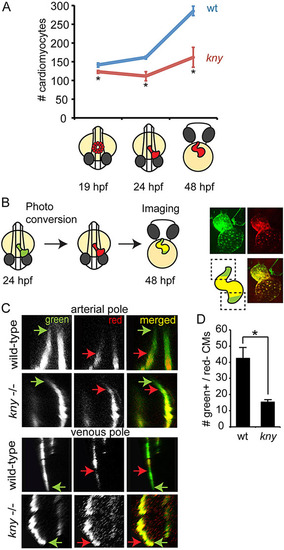
Reduced cardiomyocyte accretion in kny/gpc4 mutants. (A) Graphical representation of total cardiomyocyte counts in wild-type (wt) siblings and kny/gpc4 mutants (kny) at depicted stages. (B) Schematic of photoconversion experiment, carried out at 24hpf on Tg(myl7:nlsKikGr) or Tg(myl7:galFF;UAS:Kaede) embryos and imaged at 48hpf to visualize non-photoconverted protein (green) and photoconverted protein (red). Boxed areas indicate regions at arterial (top) and venous (bottom) poles that were imaged to analyze the accretion of cardiomyocytes (green only). (C) Optical section at the boxed areas indicated in B at the arterial or venous pole of photoconverted Tg(myl7:galFF;UAS:Kaede) wild-type siblings and kny/gpc4 mutants. Arrows in corresponding colors indicate the border of the green or the red signal, respectively. (D) Quantification of the amount of green+/red cardiomyocytes that were accreted to the arterial pole between 24 and 48hpf (n=3). Tg(myl7:nlsKikGr) was used for quantification. Results are represented as mean±s.e.m. *Pd0.05. |