
Fig. 4
- ID
- ZDB-FIG-150710-18
- Publication
- Punnamoottil et al., 2015 - Motor neuron-expressed microRNAs 218 and their enhancers are nested within introns of SLIT2/3 genes
- Other Figures
- All Figure Page
- Back to All Figure Page
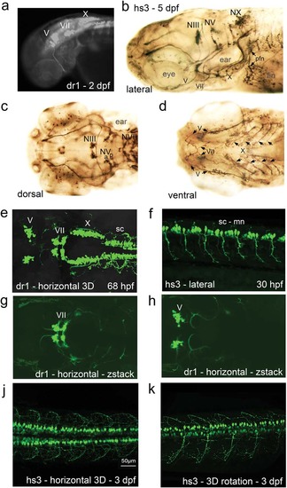
Two enhancers regulating zebrafish miR218a-1 (dr1) and human miR218-2 (hs3) drive GFP in transgenic embryos and larvae. GFP expression is found in cranial nerve nuclei and in spinal cord motor neurons and distributed in the axons. (a) Overview fluorescent microscope image. (b–d) GFP expression pattern was enhanced through anti-GFP immunostaining. The images are from different larvae and in different orientations. (e–k) Confocal live imaging. (e) z-stack image through the whole expression domain as scanned in this specimen. (f) Optical section showing spinal cord motor neurons in 30 hpf embryos. (g,h) Two z-stack images of the scan shown in “e”; one is further dorsal with focus on the facial nerve nucleus (g), while the trigeminal nerve nuclei appear further ventral in this scan (h). (j,k) 3D projections of spinal cord motor neurons. Abbreviations: hpf, hours post fertilization; mn, motor neurons; sc, spinal cord; NIII, oculomotor nerve nucleus; NV, trigeminal nerve nucleus (a, anterior; b, posterior); NVII, facial nerve nucleus; NX, vagal nerve nucleus; pfn, pectoral fin nerve. Note that V, VII, X are the corresponding nerves and X has several branches underneath the ear as seen in the lateral view. |
| Gene: | |
|---|---|
| Fish: | |
| Anatomical Terms: | |
| Stage Range: | Prim-15 to Protruding-mouth |