
Fig. 1
- ID
- ZDB-FIG-150617-1
- Publication
- Wu et al., 2015 - Hypoxia-Induced Retinal Neovascularization in Zebrafish Embryos: A Potential Model of Retinopathy of Prematurity
- Other Figures
- All Figure Page
- Back to All Figure Page
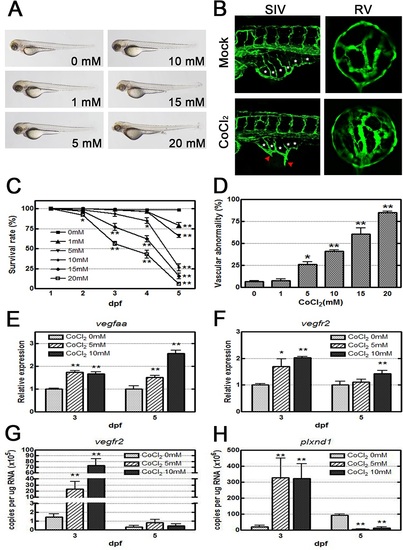
CoCl2 induces abnormal neovascularization in zebrafish embryos. (A) Morphological images obtained by an optical microscope revealed no severe phenotype in CoCl2-treated Tg(fli1a:EGFP) embryos at 3 dpf. (B) With fluorescence excitation, ectopic SIV and excessive retinal vascularization are shown in CoCl2-treated embryos compared with the untreated control. (C, D) A dose-dependent decrease in survival rates (four independent experiments; n = 35 in each group) in embryos treated with increasing concentrations (0–20 mM) and an increase in the vascular defect occurrence rate in the SIV and retinal vessels are shown (three independent experiments; n = 35 in each group). (E, F) Real-time RT-PCR data show that CoCl2 treatment causes overexpression of vegfaa and vegfr2 mRNAs in zebrafish. (G,H) Absolute quantification of the copy number of vegfr2 and plxnd1 by real-time PCR are reduced at 5 dpf versus 3 dpf. Each bar represents the mean ± SEM. * (p < 0.01) and ** (p < 0.001) compared with the mock control group. SIV, subintestinal vessel; RV, retinal vessel. |
| Gene: | |
|---|---|
| Fish: | |
| Condition: | |
| Anatomical Terms: | |
| Stage: | Protruding-mouth |
| Fish: | |
|---|---|
| Condition: | |
| Observed In: | |
| Stage: | Protruding-mouth |