
Fig. 1
- ID
- ZDB-FIG-150609-15
- Publication
- Bührdel et al., 2015 - In vivo characterization of human myofibrillar myopathy genes in zebrafish
- Other Figures
- All Figure Page
- Back to All Figure Page
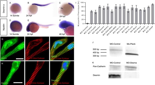
MFM disease gene expression analysis and knock-down in zebrafish (A–E) Whole-mount antisense RNA in situ hybridization shows desmin-a and myotilin expression in skeletal muscle fibers at the 14 somite stage and 24 hpf. (C,F) At 24/48 hpf cardiac expression of desmin a/myotilin was observed (arrows). (G) As revealed by co-stainings with an antibody against Z-disk Titin (T12) or DAPI, respectively, BAG3 localizes to sarcomeric Z-disks and nuclei of zebrafish cardiomyocytes. (H) Desmin is localized at the sarcomeric Z-disk, scale bars = 10 µm. (I) Percentage of injected embryos showing a myopathic phenotype. (J) Injection of MO-Plecb blocks the splice donor site of exon 7 of Plectin b, leading to an abnormally short cDNA fragment due to skipping of exon 7. (K) Western-blot analysis shows the reduction of Desmin after injection of MO-Desma. Pan-Cadherin is used as loading control. |
| Genes: | |
|---|---|
| Fish: | |
| Anatomical Terms: | |
| Stage Range: | 14-19 somites to Long-pec |