
Fig. 3
- ID
- ZDB-FIG-150603-5
- Publication
- Kaufman et al., 2015 - Development and origins of Zebrafish ocular vasculature
- Other Figures
- All Figure Page
- Back to All Figure Page
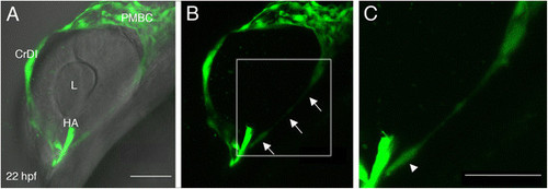
Sprouting from the ventral PMBC. (A-C) Projections of confocal z-stacks from a live kdrl:EGFP transgenic embryo at 22 hpf. In A, the same confocal image shown in B is combined with bright field image to demonstrate position of blood vessels relative to eye tissues. Arrows in B point at a long extension from the ventral PMBC towards the optic fissure, where the HA enters the eye. (C) A higher magnification of the region marked by white rectangle in B suggests the extension is actually a trail of cells. Arrowhead in C points at what appears to be an endothelial cell body. CrDI, cranial division of internal carotid artery; HA, hyaloid artery; L, lens; PMBC, Primordial midbrain channel. Anterior is to the left and dorsal up. Scale bars are 50 µm. |