
Fig. 2
- ID
- ZDB-FIG-150602-2
- Publication
- Hirose et al., 2014 - Mechanistic target of rapamycin complex 1 signaling regulates cell proliferation, cell survival, and differentiation in regenerating zebrafish fins
- Other Figures
- All Figure Page
- Back to All Figure Page
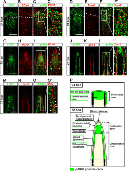
Distributions of S6K-positive cells and proliferative cells at 24 and 72 hpa. (A-F′) Longitudinal sections of wild-type fin regenerates that were co-immunohistochemically stained with antibodies against p-S6K (green) and PCNA (red) (A-C′, n = 3) or p-S6K (green) and Runx2 (red) (D-F′, n = 3) at 24 hpa. p-S6K positive cells were PCNA- or Runx2-positive at 24 hpa (C′,F′). The boxed areas in C and F are enlarged in C′ and F′, respectively. Dashed white lines indicate the amputation planes. (G-O′) Longitudinal sections of wild-type fin regenerates that were co-immunostained with antibodies against p-S6K (green) and PCNA (red) (G-I′, n = 5), p-S6K (green) and Zns-5 (red) (J-L′, n = 3), or p-S6K (green) and Runx2 (red) (M-O′, n = 4) at 72 hpa. PCNA-positive cells were p-S6K-positive in the putative proximal medial blastema domain (arrowheads in I′), but not in the tip of the putative proximal medial blastema domain (a bracket in I′). Zns-5- or Runx2-positive cells in the proximal lateral blastema were also p-S6K-positive (arrowheads in L′ and O′), but cells in the distal region of the lateral blastema were not (brackets in L′ and O′). It should be noted that both p-S6K and PCNA or Runx2 fluorescent signals did not overlap, because p-S6K and PCNA or Runx2 are localized in the cytosol and nucleus, respectively. Scale bars: 100 µm. (P) Cartoon summarizing the anatomical structures of fin regenerates in longitudinal cross-sections and localization of p-S6K-positive cells in the fin regenerates at 24 and 72 hpa. Dashed lines indicate the amputation planes. |