FIGURE
Fig. 5
- ID
- ZDB-FIG-150527-16
- Publication
- Meyen et al., 2015 - Dynamic filopodia are required for chemokine-dependent intracellular polarization during guided cell migration in vivo
- Other Figures
- All Figure Page
- Back to All Figure Page
Fig. 5
|
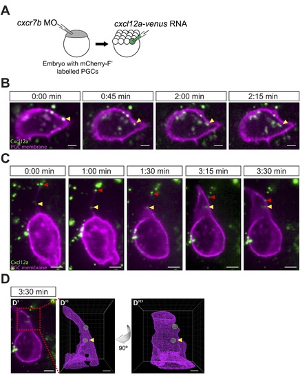
Cxcl12a internalization and interaction with filopodia. (A) Schematic experimental setup. Cxcr7b function was knocked down in embryos, in which PGCs express mCherry on their membrane. At 16-cell stage, these embryos were injected with cxcl12a-venus RNA directed into a corner cell for a mosaic expression of the chemokine. (B) Cxcl12a (green) is bound to the PGC membrane (magenta) and internalizes into the cell. Snapshots from Video 8, showing an optical section of a PGC (a Z-projection of two 1-µm-slices). An arrowhead points at an internalizing Cxcl12a spot. Scale bar is 5 µm. (C) Snapshots from Video 9, showing an optical section of a PGC (a Z-projection of 4 1-µm-slices) with Cxcl12a (green) interaction seen on the filopodium (magenta). The red arrowhead indicates a Cxcl12a spot bound to the tip of the retracting filopodium and the yellow arrowhead points at Cxcl12a, which is bound to the filopodium closer to the cell body and is then engulfed by the cell (1:30–3:30 min). Scale bar is 5 µm. (D) PGC from panel C at 3:30 min (D′) where the area magnified in D3 and D4 is delineated in a red box. (D′′ and D′′′) A 3D wire presentation of the magnified cell surface area (magenta) and the relevant Cxcl12 foci (green). (D′′) A 3D image orientated as the original panel in C and (D′′′) is horizontally rotated (90° clockwise) to visualize internalization of Cxcl12a. Scale bar is 2 µm. |
Expression Data
Expression Detail
Antibody Labeling
Phenotype Data
Phenotype Detail
Acknowledgments
This image is the copyrighted work of the attributed author or publisher, and
ZFIN has permission only to display this image to its users.
Additional permissions should be obtained from the applicable author or publisher of the image.
Full text @ Elife

