
FIGURE
Fig. 3
- ID
- ZDB-FIG-150428-10
- Publication
- Luz et al., 2013 - Fluorescently tagged Lin7c is a dynamic marker for polarity maturation in the zebrafish retinal epithelium
- Other Figures
- All Figure Page
- Back to All Figure Page
Fig. 3
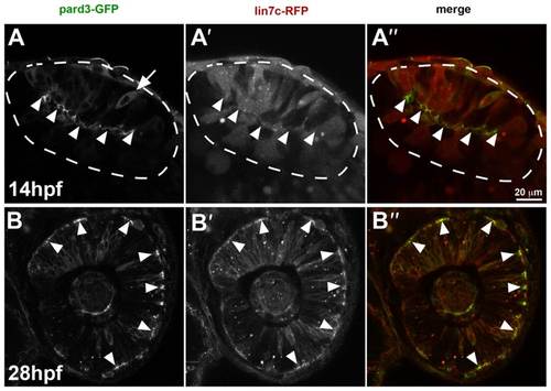
Lin7c-RFP subcellular localisation reflects temporal changes in retinal neuroepithelial polarity. (A–A′′) In the optic vesicle (14 hpf, 10ss), Pard3-GFP is observed predominantly at the apical surface (arrowheads) of cells, except for a few cells, which show a cytoplasmic distribution (arrow) (A). Lin7c-RFP is cytosolic and not observed apically (A′). (B–B′′) Cells of the retinal neuroepithelium (28 hpf) show apical localization of both Pard3-GFP (B) and Lin7c-RFP (B′). Scale bar: 20µm. |
Expression Data
Expression Detail
Antibody Labeling
Phenotype Data
Phenotype Detail
Acknowledgments
This image is the copyrighted work of the attributed author or publisher, and
ZFIN has permission only to display this image to its users.
Additional permissions should be obtained from the applicable author or publisher of the image.
Full text @ Biol. Open