
FIGURE
Fig. S2
Fig. S2
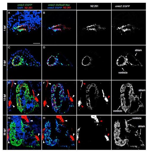
N2.261 antibody reacts with the embryo-specific cardiac myosin heavy chain isoform (embCMHC). (A-H) Heart sections of double transgenic fish cmlc2::EGFP; cmlc2::ds-Red-Nuc at 1, 3, 12 and 30 days post fertilization (dpf). Before the immunostaining, the endogenous signal was quenched by 1M HCL treatment for 30 min followed by antibody staining against GFP, DsRed and N2.261.At 1 dpf (A-B), N2.261 detects all CMs of the primordial heart. At 3 dpf (C-D), N2.261 labels the embryonic ventricle, but not atrium. At 12 dpf (E-F), the N2.261 reactivity is confined to a subset of CMs located in the middle of the ventricle. No expression is detected at the outer cell layer of the ventricular wall. At 30 dpf (G-H), the antigen of N2.261 is not longer expressed in the juvenile heart. Some of skeletal muscles adjacent to the heart are strongly labeled by N2.261 (arrowheads in E-H). Scale bar (A) = 50 µm. |
Expression Data
Expression Detail
Antibody Labeling
Phenotype Data
Phenotype Detail
Acknowledgments
This image is the copyrighted work of the attributed author or publisher, and
ZFIN has permission only to display this image to its users.
Additional permissions should be obtained from the applicable author or publisher of the image.
Reprinted from Developmental Biology, 399(1), Sallin, P., de Preux Charles, A., Duruz, V., Pfefferli, C., Jazwinska, A., A dual epimorphic and compensatory mode of heart regeneration in zebrafish, 27-40, Copyright (2015) with permission from Elsevier. Full text @ Dev. Biol.