
Fig. 2
- ID
- ZDB-FIG-150330-15
- Publication
- Yamanaka et al., 2015 - Rotating pigment cells exhibit an intrinsic chirality
- Other Figures
- All Figure Page
- Back to All Figure Page
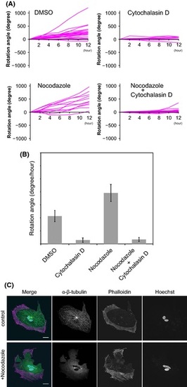
Effect of cytoskeleton inhibitors on cellular rotation. (A) The angles of rotation of melanophores treated with cytoskeleton inhibitors were observed for 12 h: DMSO (dimethyl sulfoxide) treatment (N = 24), cytochalasin D treatment (N = 10), nocodazole treatment (N = 11), and nocodazole and cytochalasin D treatment (N = 20). See Movie S3–6 (Supporting Information). (B) Effect of cytoskeleton inhibitors on cellular rotation. Cytochalasin D effectively inhibited cellular rotation, whereas nocodazole accelerated cellular rotation. The mean rates of rotation of the melanophores were as follows: DMSO (dimethyl sulfoxide) treatment (21.4°/h, SEM ± 4.3°, N = 24), cytochalasin D treatment (2.9°/h, SEM ± 1.7°, N = 10), nocodazole treatment (39.1°/h, SEM ± 6.6°, N = 11), and nocodazole plus cytochalasin D treatment (3.4°/h, SEM ± 1.5°, N = 11). (C) The actin cytoskeleton and microtubule network of rotating melanophores. The control melanophores were round, with actin fibers localized in the peripheral region and microtubules radially extended from the central region (control). When microtubule formation was inhibited by nocodazole, the actin fibers accumulated in certain sites in the peripheral region. The melanophores became irregularly shaped (+nocodazole). Scale bars represent 20 µm. |